How Does the Heart Respond?
The heart is a siphon, typically thumping around 60 to 100 times each moment. With every heartbeat, the heart sends blood all through our bodies, conveying oxygen to each cell. In the wake of conveying the oxygen, the blood gets back to the heart. The heart at that point sends the blood to the lungs to get more oxygen. This cycle rehashes again and again.
How Does the Circulatory System Respond?
The circulatory framework is comprised of veins that divert blood from and towards the heart. Conduits divert blood from the heart and veins convey blood back to the heart.
The circulatory framework conveys oxygen, supplements, and chemicals to cells, and eliminates side-effects, similar to carbon dioxide. These streets travel one way in particular, to keep things going where they ought to.
What Are the Parts of the Heart?
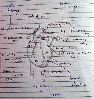
The heart has four chambers — two on top and two on base:
The two base chambers are the correct ventricle and the left ventricle. These siphon blood out of the heart. A divider called the interventricular septum is between the two ventricles.
The two top chambers are the correct chamber and the left chamber. They get the blood entering the heart. A divider called the interatrial septum is between the atria.
Representation: Healthy Heart
The atria are isolated from the ventricles by the atrioventricular valves:
The tricuspid valve isolates the correct chamber from the correct ventricle.
The mitral valve isolates the left chamber from the left ventricle.
Two valves likewise separate the ventricles from the huge veins that convey blood leaving the heart:
The pulmonic valve is between the correct ventricle and the aspiratory corridor, which conveys blood to the lungs.
The aortic valve is between the left ventricle and the aorta, which conveys blood to the body.
What Are the Parts of the Circulatory System?
Two pathways come from the heart:
The aspiratory flow is a short circle from the heart to the lungs and back once more.
The foundational dissemination conveys blood from the heart to the wide range of various pieces of the body and back once more.
In aspiratory dissemination:
The aspiratory vein is a major conduit that comes from the heart. It parts into two principle branches, and carries blood from the heart to the lungs. At the lungs, the blood gets oxygen and drops off carbon dioxide. The blood at that point gets back to the heart through the pneumonic veins.
In foundational dissemination:
Then, blood that profits to the heart has gotten loads of oxygen from the lungs. So it would now be able to go out to the body. The aorta is a major corridor that leaves the heart conveying this oxygenated blood. Branches off of the aorta send blood to the muscles of the actual heart, just as any remaining pieces of the body. Like a tree, the branches gets more modest and more modest as they get farther from the aorta.
At each body section, an organization of minuscule veins called vessels associates the tiny corridor branches to little veins. The vessels have extremely dainty dividers, and through them, supplements and oxygen are conveyed to the cells. Side-effects are brought into the vessels.
Vessels at that point lead into little veins. Little veins lead to bigger and bigger veins as the blood moves toward the heart. Valves in the veins keep blood streaming the right way. Two huge veins that lead into the heart are the unrivaled vena cava and second rate vena cava. (The terms prevalent and mediocre don't imply that one vein is superior to the next, yet that they're situated above and underneath the heart.)
When the blood is back in the heart, it needs to return the aspiratory dissemination and return to the lungs to drop off the carbon dioxide and get more oxygen.
How Does the Heart Beat?
The heart gets messages from the body that reveal to it when to siphon pretty much blood contingent upon an individual's requirements. For instance, when we're dozing, it siphons barely enough to accommodate the lower measures of oxygen required by our bodies very still. Yet, when we're working out, the heart siphons quicker with the goal that our muscles get more oxygen and can work more diligently.
How the heart beats is constrained by an arrangement of electrical signs in the heart. The sinus (or sinoatrial) hub is a little space of tissue in the mass of the correct chamber. It conveys an electrical sign to begin the contracting (siphoning) of the heart muscle. This hub is known as the pacemaker of the heart since it sets the pace of the heartbeat and makes the remainder of the heart contract in its musicality.
These electrical motivations make the atria contract first. At that point the motivations head out down to the atrioventricular (or AV) hub, which goes about as a sort of transfer station. From here, the electrical sign goes through the privilege and left ventricles, making them contract.
One complete heartbeat is comprised of two stages:
The primary stage is called systole (SISS-tuh-lee). This is the point at which the ventricles agreement and siphon blood into the aorta and pneumonic corridor. During systole, the atrioventricular valves close, making the main sound (the lub) of a heartbeat. At the point when the atrioventricular valves close, it holds the blood back from returning up into the atria. During this time, the aortic and pneumonic valves are available to permit blood into the aorta and aspiratory corridor. At the point when the ventricles wrap up getting, the aortic and aspiratory valves near keep blood from streaming once again into the ventricles. These valves shutting is the thing that makes the subsequent sound (the name) of a heartbeat.
The subsequent stage is called diastole (bite the dust AS-tuh-lee). This is the point at which the atrioventricular valves open and the ventricles unwind. This permits the ventricles to load up with blood from the atria, and prepare for the following heartbeat.
How Might I Help Keep My Child's Heart Healthy?
To help keep your youngster's heart sound:
Empower a lot of activity.
Offer a nutritious eating routine.
Help your kid reach and keep a solid weight.
Go for ordinary clinical exams.
Educate the specialist concerning any family background of heart issues.
Function Of Heart
About Me

- Priyanshu Parmar
- Himmtanagr, Gujarat, India
Search This Blog
Types of Post
Popular Posts

Nursing Diagnosis.
5:43 PM

Communication In Nursing
4:17 PM
discription
Nursing education,Nursing education pamphlet,Nursing education ppt,Nursing education pdf,Nursing education picture.
Recent Posts
3/recent/post-list
Subjects
Created with by OmTemplates | Distributed By Gooyaabi Templates


